Category Archives: Eye Services

Chalazion Surgery

What is a chalazion?

A chalazion or chalazion is a small cyst or nodule, typically between 2 and 8 mm, filled with fluid, with the appearance of a “bump or lump on the eyelid” (see photo above). It can develop on the lower eyelid or the upper eyelid, although it is more common on the upper eyelid. Chalazion can affect just one eye or both eyes.

To better identify what a chalazion is in the eye, see the images or photos above, where a lump in the upper eyelid is clearly identified. The chalazion can develop on the outer face of the eyelid (external chalazion) or on the inner face (internal chalazion) and may even grow over the free extremity (marginal chalazion).

Chalazion in the eye is a frequent disease and often disappears spontaneously (without any intervention). It is not a malignant disease and it is not contagious. Generally speaking, chalazion in the eye is not dangerous and does not affect vision, as the lump is not large enough to distort the ocular surface and cause reduced visual acuity. However, under certain circumstances vision can be affected if the swelling is considerable.

Although sometimes confused, hordeolum (inflamed eyelid gland) and chalazion are distinct diseases.

Causes of chalazion

The causes for the appearance of chalazion are all those that prevent the drainage of fat from the Meibomian glands. If the gland becomes blocked, then the fluid cannot be released and it produces a fluid-filled “bump” (cyst) forming the chalazion. Some people have thicker meibomian gland secretions, so they are more likely to develop a chalazion. Blepharitis can also be the cause for the appearance of chalazion.

Some skin conditions such as eczema and acne rosacea are also one of possible causes of chalazion. In rare cases, chalazia can be caused by skin cancer.

If a patient has already developed a chalazion in the past, there is an increased risk of developing the disease again.

In terms of prevention, hand and face hygiene, as well as avoiding contact with the hands, fundamentally, when they are dirty with the eyes is one of the main measures to be taken, as dirt increases the risk of developing the disease. The repeated appearance of chalazions may indicate a refractive problem, such as astigmatism, myopia or hyperopia, which is why an appointment with an ophthalmologist is of paramount importance to diagnose possible refractive errors or other vision problems.

Chalazion signs and symptoms

In chalazion, the following signs and symptoms are usually evident:

chalazion external

  • swelling or lump in the eyelid;
  • photophobia (sensitivity to light);
  • blurred vision;
  • decrease in palpebral fissure.

See in the first photo a chalazion that has developed on the outer surface of the eyelid (external chalazion).

Chalazion can also develop internally.

chalazion internal
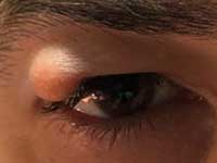
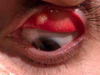
See in the second photo a chalazion on the inner face (internal chalazion).

Is there a cure for chalazion?

The chalazion has a cure. If the chalazion in the eye does not disappear spontaneously, usually within a month, surgery is required to resolve the problem. See below how to treat the chalazion.

Treatment for chalazion

In chalazion, medical treatment usually includes the administration of anti-inflammatory drugs in drops (eye drops) or ointment.

Some patients who recurrently develop chalazion may benefit from the administration of low-dose oral tetracycline, which alters the metabolism of the Meibomian glands.

Chalazion surgery

Chalazion operation or surgery is usually the last resort in treating the condition. Chalazion surgery or exeresis consists of a small surgical intervention. Usually, the operation is performed under local anesthesia which lasts between 5 to 10 minutes. Chalazion surgery does not leave a scar, as it is usually performed on the inside of the eyelid.

Chalazion surgery recovery

In chalazion surgery, recovery is fast. The recovery time is short, and patients can return to their normal activities one day after surgery.

How much does surgery cost?

The price of a chalazion surgery may vary according to the type of procedure required and the associated health insurance (if any). However, in most cases, the operation is relatively simple allowing its value not to be high.

Only the ophthalmologist will be able to know how much a chalazion surgery costs, after evaluation in consultation.

pterygium surgery

Pterygium Surgery

What is a pterygium?

Pterygium is a degeneration of the limbic conjunctiva that grows over the surface of the cornea towards the pupil. For this reason, patients often refer to pterygium as a kind of “grown flesh in the eyes” or simply, “flesh in the eyes”.

Pterygium in the eye is a lesion that usually grows slowly throughout life. It may even stop growing at a certain point. In advanced cases, the pterygium can grow so large that it covers the pupil and interferes with vision.

Ocular pterygium can vary from a small lesion to large, aggressive or fibrovascular lesions that can distort the topography of the cornea and in advanced cases can occlude the optic center of the cornea.

It is a disease that usually affects people who spend a lot of time outdoors, exposed to wind and sun, and can affect one or both eyes (bilateral pterygium).

Pterygium is usually not a serious problem. It can, however, cause some symptoms and quite unpleasant signs.

Symptoms in Pterygium

In pterygium, usually, there are several signs and symptoms that can be more or less exuberant according to the evolution of the disease. In certain circumstances, pterygium is even asymptomatic (without symptoms). Among the most common symptoms we can include:

  • Foreign body sensation in the eye;
  • Blurred vision;
  • Itchy eye;
  • Burning sensation.

A pterygium in an advanced stage can cause red eyes and inflammation in the eye.

The pterygium can progressively grow over the cornea (the clear, outer layer of the eye), and can distort the cornea, causing astigmatism.

Causes of pterygium

The causes for the appearance of pterygium can be the following:

  • Prolonged exposure to ultraviolet light (sunlight without sunglasses)
  • Dry eye;
  • Irritants such as dust and wind.

Pterygium is most common in young adults between the ages of 20 and 40 and appears to be more common in men than women. Pterygium in the eye is often preceded by another degeneration of the conjunctiva called a pinguecula. Pinguecula is a kind of yellowish neoformation in the conjunctiva near the cornea and presents the same risk factors as pterygium, especially frequent exposure to the sun without sunglasses. Since the pinguecula can prevent tears from covering the ocular surface evenly, it can cause dry eye and foreign body sensation in the eye. The pinguecula may also become reddish.

As for pterygium prevention, sunglasses can provide the best protection against ultraviolet light, dust, and wind. It’s important to protect your eyes while driving too, because, unlike the front windshield, most car side windows do not provide adequate UV protection. Even on cloudy days, ultraviolet light can penetrate through clouds. Choose sunglasses that offer good protection against ultraviolet rays.

Pterygium treatment

Pterygium usually does not require treatment if the symptoms are moderate. If there is a temporary aggravation of the problem, we must treat the pterygium with anti-inflammatory eye drops.

In certain circumstances, surgical treatment is necessary. See below under what conditions we should resort to surgical treatment of the disease.

Pterygium surgery

The use of pterygium surgery should only be considered in certain circumstances. Surgery consists of removal or excision of the pterygium.

The pterygium operation should be considered in the following circumstances:

  • Conservative treatments failed;
  • The patient’s visual acuity is compromised;
  • The patient is uncomfortable with the aesthetic appearance.

Surgery sometimes uses the patient’s own conjunctiva or preserved amniotic membrane (placenta) to fill in the empty space created by removing the pterygium. In this procedure the pterygium is removed and the amniotic membrane or conjunctiva is placed and sutured in the affected area.

Pterygium surgery with conjunctiva or amniotic membrane transplantation is usually performed under local anesthesia and takes about 30 minutes.

Pterygium surgery – risks, complications

In pterygium surgery, among the main risks and complications is recurrence after removal or excision of the pterygium.

Without a conjunctival or amniotic graft, the rate of recurrent pterygium can be high (about 30%). Removing a pterygium with a conjunctival or amniotic graft is associated with a lower risk of recurrence, which can fluctuate between 5% and 10%.

Surgical complications can also include scarring of the cornea and perforation of the white part of the eye. In some cases, surgical removal of pterygium can induce astigmatism.

Post-operative in pterygium surgery

In pterygium surgery, the postoperative period is not painful. Patients are often advised to use steroid eye drops for several weeks or months, which may reduce inflammation and the likelihood of the pterygium coming back.

Patients need to be carefully followed for one year. About 97% of recurrences occur during the first 12 months after surgery.

Recovery from pterygium surgery

In pterygium surgery, the recovery and rest time is short, and patients can resume work or normal activities a few days after the operation.

However, some additional care is needed in the first days after pterygium surgery. Patients generally need to wear an occluder in the operated eye during the first two days (48 hours) in order to prevent possible risks of infection.

How much does surgery cost?

The price of pterygium surgery may vary according to the surgical technique, and the health insurance, among other factors.

After assessing the patient in an outpatient consultation, the Ophthalmologist will be able to define the surgical plan and the cost of the surgery.

Comprehensive Eye Checkup

A comprehensive eye checkup is a thorough evaluation of the health and function of your eyes. It is important to have regular eye exams to detect any vision or health problems early on and to ensure that your eyes are functioning properly. Here is what you can expect during a comprehensive eye checkup:

Vision test: This is the most common test during an eye exam. It involves reading from a chart or looking at a computer screen to determine your visual acuity or the clarity of your vision. You will be asked to read letters or numbers of different sizes to determine your visual acuity at different distances.

Eye muscle test: This test checks the movement and alignment of your eyes. The doctor will ask you to follow a moving object or light with your eyes and will assess your eye muscles to see if they are working properly.

Pupil dilation: During this test, your pupils will be dilated, or widened, with drops to allow the doctor to get a better view of the inside of your eye. Dilation is important because it allows the doctor to see the retina, optic nerve, and other structures in the back of the eye. It can also help to detect certain eye conditions such as glaucoma, cataracts, and age-related macular degeneration.

Refraction test: This test determines your eyeglass or contact lens prescription. You will be asked to look through a machine called a phoropter and will be shown a series of lenses of different strengths. The doctor will ask you which lens gives you the clearest vision and will use that information to determine your prescription.

Ocular health evaluation: The doctor will examine the overall health of your eyes, including the front and back of the eye. This may involve checking the pressure inside your eye, examining the blood vessels in the back of your eye, and looking for any signs of infection or disease.

Glaucoma test: Glaucoma is a condition that damages the optic nerve and can cause vision loss. The doctor may use a special tool to measure the pressure inside your eye or may use a machine to take pictures of the inside of your eye to check for signs of glaucoma.

It is important to have a comprehensive eye checkup at least once every two years, or more frequently if you have a family history of eye disease or if you are at risk for certain conditions such as diabetes or high blood pressure. Regular eye exams can help to ensure that your eyes are healthy and that any problems are detected and treated early on. So, it is very important to have a comprehensive eye checkup for maintaining good eye health.

dry eye

Dry Eye

dry eye

What is dry eye?

We say that we are facing a dry eye when there are not enough tears in quantity and quality to keep the eyes lubricated, and the problem can affect only one eye (unilateral) or, then, both eyes (bilateral). In most cases, the problem affects both eyes.

On certain occasions or circumstances, the eye can be dry without any eye problems or eye disease. Pollution, excessive exposure to the sun, wind, exposure to air conditioning or environments that somehow make the air drier are factors that can facilitate the evaporation of tears and, therefore, cause dry eyes.

Watching television for long periods or frequent use of computers, cell phones, tablets, and other digital devices also causes dry eyes. This fact is related to the difference between the number of times we “blink” our eyes in a usual way and when we use this type of devices. In addition to these, other factors may be at the origin of the problem. See more information on factors that contribute to dry eye and prevention.

In some patients, there may be an eye problem (pathology) that causes dry eye, as we will see below.

Dry eye syndrome

In dry eye syndrome or dry eye disease, there is not enough tear production or, then, the tears, despite being sufficient, do not have the necessary quality to keep the eyes lubricated.

Tears also aim to protect the ocular surface from infections and harmful effects of the environment. These contain vitamins, minerals, proteins and lipids. The presence of all these substances makes it possible to obtain quality tears to hydrate, nourish and lubricate the cornea.

Tears are produced by the lacrimal glands, located behind the upper eyelid. With blinking, they spread out and make the corneal surface shiny and transparent, forming the tear film.

The tear film is made up of three layers: lipid, aqueous, and mucin. The lipid layer is the outermost and prevents tear evaporation, the innermost layer is the mucin and allows the tear film to adhere to the cornea, in turn, the thickest layer is the aqueous layer and consists of water.

The eye can be considered dry not only because of the lack of sufficient amounts of these constituents, but also when the quality is altered. The eye can be dry not only because of the quantity of these constituents but also if the quality is altered.

Thus, if the diagnosis of dry eye syndrome is confirmed, as the patient has poor ocular lubrication, the eye suffers a kind of aggression, even trauma, with blinking.

When dry eyes occur when waking up, this problem is probably related to the dry eye syndrome or disease, that is, there is a decrease in the amount of tears, essentially in the basal production, or a deficiency in their quality, namely, alteration of the lipid layer or mucin from the tear film.

Dry eye symptoms

In dry eyes, one of the most common symptoms is redness and burning or itching in the eyes. Excessive production of tears (“ watery eyes ”), excessive irritation of the eye (from smoke or wind) and discomfort and complications with the use of contact lenses are also the most frequent signs and symptoms of dry eyes.

Patients typically report that their eyes feel dry and red, accompanied by burning. Blurred or “blurred” vision may occur, mainly at the end of the day. In some situations, there may be pain in the eyes that can be more intense in the most severe cases.

Patients suffering from dry eye syndrome experience several signs and symptoms that can cause a lot of discomforts, mainly in the presence of severe dry eye.

Causes of dry eye

The most frequent causes of dry eye are: age, menopause, continuous or excessive computer use, air conditioning, use of contact lenses and some medications (diuretics, antihistamines, benzodiazepines, antidepressants, analgesics, contraceptives, in some ocular diseases and in some systemic diseases, etc.). Dry eye can occur in people of both sexes and at any age, although females are most affected.

The diagnosis should be made by an ophthalmologist through tests that measure the quality and production of tears. The tests are quick, painless and non-invasive.

Is dry eye curable?

Dry eye syndrome has no cure. However, there are effective ways of controlling the disease that allows patients to restore perfectly normal life. Find out how to treat dry eye below.

Dry eye treatment

Treatment for dry eye essentially involves the use of artificial tears in drops (eye drops) placed in the eyes several times a day, depending on the need. Sometimes, the use of eye drops is not enough and it may be necessary to use anti-inflammatories, and antibiotics, among others, for effective control of the disease. In certain situations, the occlusion of the lacrimal points is useful, preventing the drainage of tears through the lacrimal pathways, and allowing them to remain on the ocular surface.

The ophthalmologist, based on the observation and tests carried out, will decide on the most appropriate treatment for dry eye for each patient.

Patients, in addition to medical treatment, should avoid contact with the risk factors mentioned above, such as prolonged and excessive use of the computer (taking breaks for example), avoiding air conditioning, etc.

Dry eye drops

In the treatment of dry eye, several types of artificial tears (eye drops) are used, and it is important to choose the best eye drops for an effective treatment. It is essential that these eye drops do not contain preservatives, as their use can cause serious eye allergies (conjunctivitis).

Advice from an ophthalmologist on the correct use of eye drops for dry eyes, or another medication is essential for effective treatment.

Dry eyes – what to do?

In addition to consulting an ophthalmologist in order to make a possible diagnosis of dry eye syndrome, it is also necessary to take some preventive measures or “natural treatment” for the health of your eyes.

Changing habits when using the computer or other digital devices, choosing the monitor, adapting the lighting in the workplace and at home, and routines during the day or night, among others, are all measures that can help prevent or treat the problem of dry eyes.

Keratoconus

Keratoconus

Keratoconus

What is Keratoconus?

Keratoconus is a non – inflammatory, degenerative disease of the cornea. Changes in the structure of the cornea make it thinner, modifying its curvature, thus giving it a more conical shape (ectasia) than normal (see photos above).

To better understand what keratoconus is in the eyes, look at the figure above. Look at the image on the left and see the typical curvature of a normal cornea. As you can see in the eye with keratoconus, the curvature of the cornea is modified and consequently, the images, when passing through the cornea, are distorted, causing changes in the vision of people with keratoconus (image on the right). Corneal changes induce myopia and myopic astigmatism. Myopia and astigmatism are two types of refractive errors.

Ocular keratoconus is the most common corneal dystrophy. It is a disease that affects one individual in every thousand. This disease occurs in populations all over the world, however, some ethnic groups have a higher prevalence than others. It is usually diagnosed in adolescent patients (2nd decade of life) with the most severe stage in the second and third decades of life.

The disease can affect just one eye or both eyes. Bilateral keratoconus (in both eyes) is more common than unilateral keratoconus (keratoconus in only one eye).

Keratoconus is a serious disease as it progresses to significant loss of visual acuity. In advanced stages, the patient may even go blind.

Symptoms of keratoconus

The most common symptoms of keratoconus are the perception of multiple ghost images (monocular polyopia). These symptoms are more evident in high-contrast fields of view. The keratoconus patient sees many dots scattered in a very irregular pattern. This pattern usually does not change, but it can take on new forms over time. In some cases, monocular diplopia may be present (presence of double instead of multiple images). Keratoconus often causes headaches, due to the strain on the eyes that the patient has to do to see.

Usually, keratoconus causes substantial changes in vision such as: hypovision (low vision), diplopia (perception of two images) or polyopia (multiple images) and exaggerated sensitivity to light (photophobia).

Symptoms usually worsen as the disease progresses. Keratoconus can blind, that is, it can lead to blindness in the advanced stages of the disease.

Keratoconus Diagnosis

The diagnosis of keratoconus is made by clinical observation and through complementary diagnostic tests. The most used examination in the diagnosis of keratoconus is corneal topography. Corneal topography is an eye exam that allows the evaluation of gross or incipient anomalies on the anterior or posterior surface of the cornea, allowing the diagnosis of keratoconus and other pathologies to be made.

Causes of keratoconus

The causes of keratoconus are still not completely understood. It is known that “rubbing or scratching the eyes” can help with the evolution of keratoconus, so it is important to avoid these gestures. It is also known that, usually, keratoconus evolves during pregnancy.

Degrees of keratoconus

We can identify the following degrees of keratoconus:

Grade 1 – incipient keratoconus is the first degree of keratoconus. At this stage, vision is preserved and refractive errors (myopia and myopic astigmatism) are small.

Grade 2 – grade 2 keratoconus is the next phase in which the patient already reports some hypovision and needs glasses or contact lenses to correct the resulting refractive errors.

Grade 3 – grade 3 keratoconus is an advanced stage of the disease in which vision, even with optical correction, is already quite affected, and may require surgical correction, namely, intrastromal rings.

Grade 4 – Grade 4 keratoconus is the most advanced stage where we can find leukomas (corneal opacity) or edema (hydrops). The only form of treatment is corneal transplantation.

Does Keratoconus have a cure?

There is no cure for keratoconus, however, if treated properly it can restore good visual acuity compatible with the needs of daily tasks.

Keratoconus can and should be operated on, adjusting the surgical technique to the type and stage of keratoconus. When glasses or contact lenses do not correct visual acuity to acceptable values ​​and before the cornea becomes cloudy (hydrops), the currently most accepted technique is the implantation of rings in the thickness of the cornea, according to the type and stage of keratoconus. The results are extremely encouraging.

In extreme situations of hydrops, the surgical method of choice is corneal transplantation.

Keratoconus treatment

The two forms, most used in the treatment of keratoconus, are:

1st Treat the hypovision caused by keratoconus through glasses in the initial phase of the disease and when the use of glasses does not produce the desired effects, the use of contact lenses for keratoconus should be tried, namely, semi-rigid or rigid contact lenses.

Contact lenses for keratoconus should be semi-rigid or rigid depending on the stage (incipient or advanced keratoconus), type of keratoconus and the adaptability of the patient. Each patient has a different sensitivity to adapt to contact lenses. While for one person adaptation may be easy, for another, adaptation may prove to be quite complicated.

2nd Another form of keratoconus treatment is to treat the pathology, that is, resorting to surgery, namely, by placing corneal rings in the corneal stroma in order to modify its curvature, thus obtaining excellent visual results. When the cornea has no physical conditions or is very conical or cloudy, what is indicated is a corneal transplant, in order to restore the transparency of the optical means.

Corneal ring surgery is usually performed with two types: Paulo Ferrara rings (Ferrara ring) and INTACS.

Ferrara ring and INTACS

As we have seen, corneal ring surgery can be performed using Paulo Ferrara rings (ferrara ring implant) or by INTACS.

The difference between the two methods is that the former has a smaller radius of curvature, that is, they are smaller than INTACS.

The use of one or the other will be defined by the experience of the ophthalmologist, and by the conditions and location of the keratoconus, since it can be central, inferotemporal (the most frequent) or superior (the least frequent).

To learn more about corneal ring placement surgery, see keratoconus surgery and learn about the main advantages of this surgery when compared to corneal transplantation.

Corneal Crosslinking

Currently, there are new treatments for keratoconus, with corneal crosslinking being one of those that have shown the best results, allowing to an increase in the rigidity of the cornea.

This keratoconus treatment is performed using a type of laser (UV light) on a cornea that has been previously showered with riboflavin (vitamin B2).

Most of the cornea is made up of type IV collagen fibers. These fibers are grouped in parallel and perpendicular layers. Riboflavin is a compound that is present in our body and absorbs UV light. By applying riboflavin to the cornea, and associating it with UV light, we managed to increase the rigidity of the cornea, stabilizing ectasia.

Over time, the cornea becomes progressively more rigid (Crosslinking).

Keratoconus surgery

Keratoconus surgery can be performed by placing corneal rings in the corneal stroma in order to correct its curvature. Keratoconus surgery with implantation of an intrastromal ring or corneal rings has lower intra and postoperative risks and complications, and faster recovery, among other advantages, when compared with corneal transplantation. In patients with keratoconus, the surgical results obtained with intrastromal ring implantation are very good and usually better than those achieved with corneal transplantation, for several reasons:

  • Visual acuity after corneal rings is usually higher than expected in corneal transplantation;
  • Refractive errors after an intracorneal ring are much smaller than after corneal transplantation and recovery is much faster;
  • Ring surgery is reversible, whereas transplant surgery is irreversible, and corneal decompensation may occur after a few weeks or after 20 years, for example;
  • Surgery for keratoconus with corneal rings is easier than transplantation, it can be performed under local anesthesia and recovery is much shorter, although it is not free of complications or intra- or postoperative risks;
  • Corneal ring surgery has advantages over transplantation even if we consider the ages of these patients, who are generally young people, not subject to corneal decompensation;
  • Ring surgery is an extra-ocular surgery, while corneal transplantation is open surgery, subject to some serious complications, namely, choroidal haemorrhage, retinal detachment, endocular infection (endophthalmitis), secondary cataracts, secondary glaucoma, synechiae of the iris, pupillary irregularity, cystoid macular edema, etc.
  • Although the results of surgery with rings are encouraging, this result is only possible with a lot of experience, a good anamnesis, good and adequate complementary diagnostic exams, a good surgical plan and a perfect surgical technique, free from errors;
  • It should be noted that the results achieved with the placement of an intrastromal ring are good, although there may be some post-surgical complications, namely, ring extrusion in traumatic situations or corneal ulcers.

When there are no conditions for this operation, what is indicated is a corneal transplant.

How much does surgery cost?

The price of keratoconus surgery may vary according to the surgical technique, subsystem or health insurance associated with the patient, etc.

After evaluating the patient in the consultation, the Ophthalmologist will be able to define the surgical plan and estimate the cost of the surgical intervention.

Hyperopia

Hyperopia

What is hyperopia?

Hyperopia is a refractive error in which images form behind the retina. To better understand the meaning of ocular hypermetropia, imagine the image of a given object passing through the eye. This image is focused so as to converge to a point located at a given distance. With normal vision, this point of convergence, where the image is focused, is the retina. That is, in a normal eye the image must form on the retina. In turn, the retina transmits information to the brain via the optic nerve.

In the eye with farsightedness, or farsightedness, the image is not formed correctly on the retina – but is formed from behind. Therefore, the image transmitted to the brain is not the correct image (see photos above).

The patient sees poorly near and far and for the images to be clear, he makes a great eye effort.

Hyperopia

Symptoms of hypermetropia 

Eye fatigue (“tired eyes”) and headaches are the first symptoms of farsightedness. The effort that the patient makes to overcome the disability may be the cause of these manifestations, more frequent in the late afternoon and after work or reading.

Until the age of 35/40, the age at which the capacity for accommodation begins to decline, low hypermetropia often goes unnoticed.

Great farsightedness, on the other hand, is easily noticeable in children because it is often accompanied by accommodative strabismus, and should be corrected as soon as possible.

Therefore, it is important to know what hypermetropia is and how to correct this refractive error, as it can cause numerous symptoms that take away a lot of quality of life for patients.

Causes of hypermetropia 

Hyperopia can have two essential causes: either because the cornea has an altered curvature, that is, it is flatter than normal, or because the length of the eye is smaller than normal. Large values ​​of hyperopia are usually related to hereditary hyperopia.

Diopters in ocular hypermetropia 

There are 3 degrees of farsightedness:

  • Light, from 0 to 2 diopters;
  • Moderate, from 2 to 6 diopters;
  • High, greater than 6 diopters.

Infantile farsightedness

The term infantile or childhood hypermetropia is used whenever refractive error affects children.

The first ophthalmological consultation should take place in the first year of life, to screen for refractive errors, namely anisometropias, thus avoiding possible amblyopias.

It is estimated that 10 to 15% of school-age children have vision problems that can influence behaviour and school performance. For this reason, in children with hypermetropia, it is important for parents to be aware of any signs and symptoms reported by children.

Children who suffer from hypermetropia may be tired, sleepy and lack concentration, due to having to exercise great accommodation to focus objects on the retina. Therefore, the correction of hyperopia must be carried out as quickly as possible.

When hyperopia in children is present in only one eye, it is even more difficult to observe any alteration, as the child has absolutely normal behavior.

Most children do not know how to refer when they do not see well. He believes that the vision he has is normal because he has never seen better.

Early detection of vision problems in children is essential not only to avoid permanent visual impairment (amblyopia) but also to avoid problems not only in learning but also in the child’s development.

Is hypermetropia curable?

Unfortunately, farsightedness has no cure. However, the correction methods currently available make it possible to offer patients with farsightedness a perfectly normal life. See below how to correct, or treat, hyperopia.

Hyperopia treatment 

In hypermetropia, the treatment (correction of the refractive error) can be carried out with glasses, or contact lenses or from the age of 20, the patient can be operated through laser (LASIK). Hyperopia can also be corrected with the use of intraocular lenses depending on the anatomical conditions of the eyeball.

If the patient is over 45 years old, treatment for farsightedness can be performed by phacoemulsification with intraocular multifocal lenses allowing good visual acuity at all distances without glasses.

Lenses for farsightedness

Farsightedness can be compensated by using a lens for convergent (convex) farsightedness, which focuses the image on the retina.

These lenses are thicker in the center than in the periphery and the difference in thickness is greater the hypermetropia.

Hyperopia and myopia

Hyperopia and myopia are two types of refractive errors of the eyeball. The difference between myopia and hyperopia is the following: in myopia, objects are focused in front of the retina, while in hyperopia, images are formed behind the retina.

Hyperopia and astigmatism

Farsightedness and astigmatism can exist simultaneously (together). Astigmatism and farsightedness are two types of refractive errors. Treatment of farsightedness with astigmatism can be done with glasses, contact lenses or surgery.

Farsightedness surgery

Farsightedness surgery can be performed using a laser (LASIK) or intraocular lenses. Surgery for hyperopia allows correction of the refractive error, however, not all patients meet the necessary conditions to undergo the operation.

Farsightedness and astigmatism surgery allows you to correct both refractive errors.

Astigmatism

What is astigmatism?

Astigmatism is a refractive error in a certain axis, in which the image on the retina appears out of focus. Astigmatism is among the most frequent vision problems and it can be associated with other refractive errors or eye diseases, as we will see later.

To better understand what is astigmatism in the eyes or ocular astigmatism, look at the images or photos above. With normal vision, the image is focused on the retina and on a single point of focus. The retina plays an important role in vision, as it receives images and transmits them to the brain through the optic nerve.

In the eye with astigmatism, objects are focused on more than one point, thus distorting the vision, that is, the images are distorted when passing through the cornea and, as such, appear out of focus when projected onto the retina. Consequently, in vision with astigmatism, the images transmitted to the brain are blurred or distorted, causing blurred or “blurred” vision.

Astigmatism

Symptoms of astigmatism

People with ocular astigmatism have the following symptoms: blurred near and far vision, that is, objects that are closer or farther away are distorted. The astigmatist or person with astigmatism may experience eye fatigue (tired eyes) or headaches (headache), which are the most frequent symptoms of astigmatism.

At an early stage, astigmatism may be asymptomatic (without symptoms), however, as the disease worsens, the first signs and symptoms appear.

Causes of Astigmatism

The causes of ocular astigmatism are unknown, and refractive error is usually present from birth. Usually, the defect is in the curvature of the cornea whose shape is more oval than round. The curvature of the cornea is asymmetrical, thus blurring the vision.

Astigmatism can also be induced by certain eye diseases, such as keratoconus, and pterygium, among others.

We say that we are facing irregular astigmatism when the curvature of the cornea is very uneven. In turn, we call it regular astigmatism when there is only a difference in curvature between two perpendicular meridians of the cornea.

We are dealing with simple astigmatism when astigmatism is the only existing refractive error or with compound astigmatism when this is associated with another refractive error, such as myopia or hyperopia. That is, two combinations can occur:

  • astigmatism and myopia ;
  • astigmatism and hyperopia.

Myopic astigmatism

We say that we are facing compound myopic astigmatism when the two refractive errors exist simultaneously (astigmatism and myopia together). Astigmatism with myopia is a very common eye problem.

This visual anomaly (myopia and astigmatism together) is common in children and can have negative repercussions on school success, as it causes hypovision. If astigmatism is unilateral, early correction in children is urgent (before the age of six), as it can cause amblyopia or loss of physiological vision that may be irreversible.

Hyperopic astigmatism

Compound hypermetropic astigmatism (astigmatism and hypermetropia together) is a frequent visual problem in both sexes.

When present in children, it can lead to major irreversible vision losses (amblyopia) if not corrected in a timely manner before 6 years of age.

Childhood astigmatism

The term infantile or childhood astigmatism is used whenever refractive error affects children. The first visit to the ophthalmologist should take place in the first year of life to screen for possible refractive errors, namely anisometropias, thus avoiding possible permanent visual impairments (amblyopias).

Most children do not know how to refer when they do not see well. He believes that the vision he has is normal because he has never seen better. The early detection and treatment of vision problems in children is essential not only to avoid possible amblyopias but also to prevent problems in their development.

It is estimated that between 10 and 15% of school-age children have eye problems that can influence their performance at school.

To learn more about astigmatism in children, see more information on myopic and hyperopic astigmatism.

Is astigmatism curable?

Astigmatism has no cure. However, if appropriate measures are taken to correct the refractive error, we will be able to restore good visual acuity to patients, compatible with the needs of their daily tasks. See below how to treat or correct astigmatism.

Treatment/correction of astigmatism

The treatment for astigmatism involves correcting the refractive error. Any astigmatism can be corrected through glasses or contact lenses. Surgical correction is also possible, resorting to laser (LASIK) or operating with intraocular lenses.

The ophthalmologist is responsible for indicating the most appropriate method of astigmatism correction for a given patient, based on several factors, such as age, ability to adapt to glasses or contact lenses, astigmatism values, etc.

See below for more information on how to correct astigmatism, namely types of lenses and surgery.

Glasses for astigmatism

Ocular astigmatism can be compensated with a toric lens (cylindrical lens) that will compensate for the corneal deficiency. The lens does not have the same thickness over the entire surface and the difference is greater the higher ocular astigmatism, in order to compensate for it.

Astigmatism is corrected with glasses, approximately until the age of fifteen, since from this age on it can be compensated by contact lenses for astigmatism. As we will see below, there is also the possibility of performing surgical treatment to correct astigmatism.

Astigmatism surgery

From the age of twenty, if the refractive error is stabilized, we can perform astigmatism surgery. The purpose of the operation or surgery is to correct astigmatism, thus restoring normal vision to patients without the need for glasses or contact lenses. Surgery for astigmatism can be performed with a laser (LASIK surgery) up to 6 diopters or through an intraocular lens in the anterior or posterior chamber (if you have no indication for LASIK). Operating astigmatism is currently a very safe option although, like any surgery, there are some risks and complications that must be considered.

Astigmatism can also be treated with intraocular lenses, placed in the capsular bag in patients over 45-50 years old, as well as presbyopia and other refractive errors.

In patients who suffer from astigmatism and myopia together, surgery for astigmatism and myopia makes it possible to correct both refractive errors simultaneously. Also, the most frequently used techniques are LASIK and intraocular lenses.

Surgical results are excellent if we take into account complementary diagnostic tests, indications and contraindications for surgical techniques, based on age, values ​​and stabilization of astigmatism.

myopia

Myopia

Myopia specialists

The clinical staff of ophthalmologists at Vijovis is specialized in the evaluation, diagnosis and surgery of myopia in Faridabad. We have the most advanced devices for the identification of diseases.

In addition to high technology, we also value good service, which ranges from punctuality to total attention and care for the patient.

myopia

What is myopia?

Myopia is a condition consisting of the inability to focus on distant objects (distance blurring) and the ability to focus on close objects clearly (viewing close objects very sharply).

In myopic patients, the image is focused before reaching the retina. The ideal place would be exactly on top of the retina, neither before nor after.

What causes myopia?

The causes of myopia occur for the following reasons:

  • Axial: occurs when the patient’s eye is very elongated. As a consequence, the image of objects that enter our eyes through the pupil cannot reach the retina and ends up being focused a little before it.
  • Refractive or curvature: occurs when either the cornea or crystalline lens (natural lenses of our eyes) have high refractive power. As a result, light rays converge intensely, causing the image of objects entering our eyes to be focused before reaching the retina.
  • Environmental and genetic: studies have already proven that children’s habit of seeing things very small and very close, in motion or with low lighting (such as tablets, video games, smartphones, etc.), causes a very large accommodative effort. With this, it contributes to the development of myopia, especially if the child has someone in the family with this disorder (hereditary-genetic factor).

Myopia Treatments

One of the myopia treatments is correction through corrective glasses or contact lenses. However, it is worth mentioning that these resources are only palliative because only while they are being used do they have the power to correct myopia. Myopia will decrease, stabilize or increase regardless of whether or not corrective lenses are used.

Myopia treatment consists of prescribing corrective glasses or contact lenses (divergent or concave lenses). These two modalities mentioned above are considered palliative since they only have an effect while they are being used (when glasses or contact lenses are removed, myopia is no longer being corrected).

Myopia surgeries

The definitive myopia correction treatment consists of laser myopia surgery using LASIK or PRK refractive surgery techniques. Laser myopia surgery is considered simple, without the need for hospitalization. It consists of flattening the cornea using the laser, correcting the degree of myopia and consequently normalizing vision.

Both through the corrective lenses of glasses or contact lenses, as well as through laser myopia refractive surgery or laser myopia corrective surgery, the objective is the same: to redirect the image so that it is formed on the retina (recalling that in the myopic the image is formed before the retina, which is the reason for blurry vision).

Our Myopia Experts

For myopia surgery, you and your doctor work as a team, so you need a myopia specialist, so you can have the best possible results with a quick and safe recovery. We have a team of renowned ophthalmologists and myopia specialists with extensive clinical experience ready to assist you.

Questions about myopia

WHAT ARE THE SYMPTOMS OF MYOPIA?

The main symptoms include:

  • The vision of blurred and blurry objects in the distance, such as the difficulty of seeing the number of the bus that is arriving at the stop;
  • The ability to see close objects very clearly;
  • The need to squint or close the eyelids to get a less blurry image in the distance
  • Sensitivity to light in very bright places, headaches and frequent eyes. In addition, there may be difficulty driving at night and in some cases excessive tearing.

IS THERE MYOPIA PREVENTION?

No, unfortunately, there is no prevention for myopia. However, some habits are related to the development and increase of myopia, such as:

  • Very intense close visual activities (cell phone, tablets, video games, reading, writing);
  • Less time for children to be exposed to outdoor activities.

On the other hand, the opposite seems to be true as well. There are studies that relate children’s outdoor activities to a lower chance of myopia in adult life.

WHAT ARE THE RISKS OF MYOPIA?

Patients who have a high degree of myopia (greater than 6 degrees) have a higher risk of retinal detachment than the normal population. This occurs because as the high myopic patient has a large eye, his retina is torn and stretched since it does not follow the growth of the eye.

This is the risk of high myopic patients: having ruptures or tears in their peripheral retina and evolving with retinal detachment.

Therefore, it is mandatory to perform a retinal mapping exam in all high myopic patients at least once a year, in order to prevent these injuries and prevent retinal detachment from occurring.

If the myopic patient notices in his field of vision the presence of black opacities and dirt (floaters), flashes of light (photopsia) or a sudden black spot, he should immediately go to a specialized ophthalmological service, and his retina should be promptly evaluated. That’s because there may have been a rupture, tear or even retinal detachment.

Therefore, due to the fact that the myopic patient has a more fragile eye and consequently a greater risk of retinal detachment than the general population, this patient must avoid blows and trauma to his eye and sports that may expose him to this type at all costs. situations (MMA, boxing, soccer, karate, etc).

WHAT ARE THE RISKS OF MYOPIA SURGERY?

The risks of surgery for myopia are minimal (especially considering the great benefit it can cause). This is true as long as all preoperative exams have been carried out correctly and care has been taken in preparing the patient and the operating room environment.

The main complication of myopia correction surgery (if we can call it a complication) is the non-complete correction of the degree after surgery, resulting in hypo or hypercorrection. This problem can be solved through a retouch after a few months of the previous procedure if the surgeon considers it necessary or possible. It is important to emphasize that it is not always possible to carry out the “retouching”, which will depend on the situation of the cornea after the initial procedure.

Other types of laser surgery complications involve:

  • The risk of infection after the procedure. Therefore, the use of antibiotic eye drops soon after refractive surgery is recommended, since this risk is extremely low);
  • Problems with corneal healing in the months following refractive laser surgery. If this occurs, such a complication can be treated with the application of specific eye drops by your specialist;
  • Glare, especially when driving at night. It is worth mentioning that this symptom usually disappears in a few weeks and at the latest in months;
  • Ocular dryness in the weeks/months following refractive surgery. If this occurs, it can be corrected with the use of lubricating eye drops until the lubrication of the eye returns to normal after a few weeks or months.

It is noteworthy that all these risks can be minimized or even neutralized through a thorough preoperative evaluation. Therefore, refractive laser myopia surgery is considered in the medical literature to be a very safe and reliable surgery.

IS THERE A MINIMUM GRADE FOR MYOPIA SURGERY?

Yes, there is. It is not advisable to perform the myopia operation when the patient’s degree is equal to or less than 0.75, although there are some exceptions to this rule.

IS THERE A MINIMUM AGE FOR MYOPIA SURGERY?

Yes, laser refractive correction is indicated for patients over 18 years old.

HOW IS THE POSTOPERATIVE PERIOD OF MYOPIA SURGERY?

Recovering from myopia surgery usually takes an average of 15 days, but this may vary according to the degree of myopia before surgery, the type of procedure performed and the ability of each patient’s eyeball to heal.

During recovery, the doctor usually asks the patient to:

  • Use the prescribed antibiotic and anti-inflammatory eye drops;
  • Avoid scratching your eyes;
  • Avoid practicing some sports, such as basketball, football, diving, swimming or tennis,
  • during the 30 days following the procedure;
  • Do not use makeup between seven and 14 days;
  • Wear sunglasses in very bright environments.

Professional activities can be carried out normally, according to the patient’s recovery. To drive, the patient must feel safe to carry out this activity and it can only be done with the authorization of his doctor.

It is very common for the view to be “blurry” in the postoperative period because of inflammation in the eyeball. In the first post-surgery days, it is normal for the patient to feel constant “burning” and itching in the eye area. However, over the days, the view becomes clearer and more defined.

Lasik Eye Surgery

It is eye surgery that permanently changes the shape of the cornea (the clear covering at the front of the eye). This is done to improve vision and reduce the person’s need to wear glasses or contact lenses.

For clear vision, the cornea and lens of the eye must bend (refract) light rays properly. This allows images to focus on the retina. Otherwise, the images will be blurry.

This blurry vision is called “refractive error.” It is caused by a mismatch between the shape of the cornea (curvature) and the length of the eye.

LASIK uses an excimer laser (an ultraviolet laser) to remove a thin layer of corneal tissue. This reshapes the cornea so that light rays are clearly focused on the retina. LASIK makes the cornea thinner.

LASIK surgery is an outpatient surgical procedure. It takes 10 to 15 minutes for each eye.

The only anesthetic used is an eye drop that numbs the surface of the eye. The procedure is done while you are awake, but you will be given medicine to help you relax. LASIK surgery can be performed on one or both eyes in the same session.

To perform the procedure, a flap of corneal tissue is created. This flap is then detached so that the excimer laser can reshape the underlying corneal tissue. A hinge on the flap prevents the flap from completely separating from the cornea.

The first time LASIK surgery was performed, a special automated knife (a microkeratome) was used to cut the flap. Now a more common and safe method is to use a different type of laser (femtosecond) to create the corneal flap.

The amount of tissue to be removed with the excimer laser is calculated in advance. The surgeon will calculate this amount based on several factors including:

  • Your prescription glasses or contact lenses
  • A wavefront test, which measures how light travels through your eye
  • The shape of the surface of the cornea

Once the reformation is done, the surgeon replaces the flap and secures it. No sutures are needed. The cornea will naturally hold the flap in place.

Why the procedure is performed

LASIK surgery is most often performed on people who wear conventional or contact lenses for nearsightedness. It is sometimes used to correct farsightedness and can also correct astigmatism.

The US Food and Drug Administration (FDA) and the American Academy of Ophthalmology have developed guidelines for determining candidates for LASIK surgery:

  • You must be at least 18 years old (21 in some cases, depending on the laser used). This is because the vision of people under the age of 18 can continue to change. The young child with one highly myopic eye and one normal eye is a rare exception. The use of LASIK to correct the highly myopic eye can prevent amblyopia (lazy eye).
  • Your eyes must be healthy and your prescription stable. If you are nearsighted, you should postpone LASIK surgery until your condition has stabilized. Nearsightedness can continue to increase in some people until age 25 to late 30.
  • Your prescription must be within the range that allows LASIK correction
  • You must be in good general health. LASIK may not be recommended for people with diabetes, rheumatoid arthritis, lupus, glaucoma, herpetic eye infections, or cataracts. You should discuss this with your surgeon.

Other recommendations:

  • Weigh the risks and benefits. If you are happy wearing glasses or contact lenses, you may not want to have this surgery.
  • Make sure you have realistic expectations of the surgery.

For people with presbyopia , LASIK surgery cannot correct vision so that one eye can see both far and near. However, LASIK surgery can be done so that one eye sees far and the other sees near. This is called “monovision.” If you can adjust to this correction, it may eliminate or reduce the need for reading glasses.

In some cases, it is only necessary to operate on one eye. If your doctor thinks you qualify, ask about the pros and cons.

Risks

You should not have this procedure done if you are pregnant or nursing, as these conditions affect vision measurement values.

You should not have this procedure if you are taking certain prescription medications, such as Accutane, Cordarone, Imitrex, or predisone by mouth.

Risks may include:

  • corneal infection
  • Corneal scarring or permanent problems with the shape of the cornea, making it impossible to wear contact lenses
  • Decreased contrast sensitivity, even with 20/20 vision, objects may appear blurry or gray
  • dry eyes
  • glare or halos
  • light sensitivity
  • Problems driving at night
  • Red or pink patches on the white of the eye (broken blood vessels, usually temporary)
  • Decreased or permanent loss of vision
  • Itch

Before the procedure

A complete eye exam will be done before surgery to make sure your eyes are healthy. Other tests will be done to measure the curvature of the cornea, the size of the pupils in light and dark, the refractive error of the eyes, and the thickness of the cornea (to make sure you have enough corneal tissue left after surgery). ).

You will sign an authorization before the procedure. This confirms that you are aware of the risks, benefits, alternative options, and potential complications of the procedure.

After the procedure

After surgery:

  • You may have burning, itching, or a feeling that something is in your eye. This sensation does not last more than 6 hours in most cases.
  • A shield or patch will be placed over the eye to protect the flap. It will also help prevent rubbing or pressure on the eye until the eye has had enough time to heal (usually overnight).
  • It is very important NOT to rub the eye after LASIK surgery, so that the flap does not become dislodged or move. For the first 6 hours, keep your eye closed as much as possible.
  • The doctor may prescribe mild pain medication and a sedative.
  • Vision is usually blurry or cloudy on the day of surgery, but will improve the next day.

Call your eye doctor right away if you have severe pain or if any of your symptoms worsen before your scheduled follow-up appointment (24-48 hours after surgery).

At the first visit after surgery, the eye shield will be removed and the doctor will examine your eye and test your vision. You will be given eye drops to help prevent infection and inflammation.

Do not drive until your vision has improved enough to drive safely. Other things to avoid include:

  • Swimming
  • Bathtubs and whirlpools
  • contact sports
  • Use of lotions, creams, and eye makeup for 2 to 4 weeks after surgery

The health care provider will give you specific instructions.

Expectations (forecast)

In most people, vision will stabilize within a few days after surgery, but for some people it can take up to 3 to 6 months.

A small number of people may need another surgery because vision is over or under corrected. Although a second surgery may improve distance vision, it may not relieve other symptoms such as glare, halos, or trouble driving at night. These are common complaints after LASIK surgery, especially when using an older method. These problems should disappear by 6 months after surgery in most cases. However, a small number of people may continue to have problems with glare. Even after a second surgery, few people may still need to wear contact lenses or glasses.

Frequent Questions

Is laser refractive surgery always 100% laser?

No. The patient must be well informed about the surgical procedure. In some centers, despite being called the Lasik technique, they still use a blade in the first part of the surgery. At Vizovis we have completely abandoned the use of the blade and have replaced it with a femtosecond laser.

Is femtosecond laser refractive surgery a painful procedure?

During the intervention you can feel a sensation of touch, pressure, but not pain. In the immediate postoperative period, during the first 5 or 6 hours, a sensation of grit, stinging, tearing, and photophobia may appear. The normal thing is that the next day there is practically no discomfort.

In femtosecond laser surgery, are both eyes operated at the same time?

Yes. The patient will also find it more comfortable if the surgery is bilateral.

Is FemtoLasik surgery a forever treatment?

The effect of the femtosecond laser is permanent, but in a small percentage of patients, over time, myopia may increase again because the eye becomes longer. It is also important for this not to happen that the ophthalmologist assures the patient that her graduation is stable. However, it can be corrected again with a touch-up.

After femtosecond laser surgery, what care should I take?

It is vitally important to avoid rubbing the eyes. You must use the eye drops as prescribed. It is convenient to use glasses as a protective measure in situations with risk of trauma or inclusion of particles.

Cataract Surgery

What are cataracts?

The cataract is the progressive opacification of the crystalline lens, so as it evolves, a gradual reduction of vision occurs. Cataracts are normally associated with age, although they can be congenital, caused by trauma or see their formation accelerated in certain diseases, as well as after taking some medications.

Usually, the loss of transparency of cataracts occurs progressively and slowly, so it may not be appreciated for a long time and one can get used to a visual acuity that is not really good enough. His solution is surgical.

How do I know if I have cataracts?

Cataracts cause a progressive loss of vision both far and near, with the sensation of blurred vision and halos around lights, with increasing discomfort when exposed to high light conditions.

Types of cataracts

Subcapsular cataract

The subcapsular cataract occurs at the back of the lens. The patients most at risk of suffering from this type of cataract are those who have diabetes or take high doses of steroids.

Nuclear cataract

The nuclear cataract occurs in the central zone of the crystalline lens. This cataract is mainly associated with age.

Cortical cataract

This type of cataract occurs in the cortex of the lens. This type of cataract occurs when small white opacities around the lens begin to move towards the center of the lens in a radial manner.

Metabolic cataract

A metabolic cataract occurs as a secondary consequence of metabolic diseases that produce unique substances in the lens that accelerate its opacification.

Congenital cataract

It is called this way when the child is born with cataracts and they can be unilateral or bilateral. Its cause is mainly hereditary and cannot be prevented. It will be important to make a diagnosis for the early detection of any type of ophthalmological condition from birth.

Traumatic cataract

This type is associated with some type of eye injury, blow or bruise that can also damage other eye structures of greater or lesser severity.

Toxic cataract

It can arise from excessive abuse of drug intake or some types of toxins such as ergot, corticosteroids or paradichlorobenzene.

Radiation cataract

It can occur due to long-term exposure to a burst of ultraviolet, infrared or ionizing radiation.

Secondary cataracts

After a few months or even years, after the surgical treatment of the cataract, the posterior capsule of the crystalline lens could become opacified, and therefore, produce a reduction in both distant and near vision. It is what we call a secondary cataract. It is produced, therefore, by a migration of the epithelial cells of the lens or anterior capsule towards the posterior capsule.

Cataract treatment

The crystalline lens is the eye’s natural lens, responsible for the quality of images captured by the retina. Once damaged by the cataract, only surgical treatment will be able to reverse the condition. There are also medications and eye drops, but they are palliative measures.

It is through surgery that the patient’s visual acuity can be completely recovered. At NeoOftalmo, the surgical technique adopted is Phacoemulsification. For this, an intraocular lens (IO’S) is implanted in the place of the lens, performing its function. In addition, this technique allows you to solve other eye problems, such as:

  • Astigmatism;
  • Hyperopia;
  • Myopia;
  • Eyestrain.

The implantation of the lens is done through several calculations that analyze the path of light before the eyeball. Before implanting the lens, the damaged lens is removed. Then the prosthesis is placed through an incision. The procedure is quite simple and does not usually cause complications.

Choosing the Intraocular Lens is a very important step!

In some cases, choosing the correct lens makes it possible to reduce the degree of the lens of the patient’s glasses or even suspend the use of the accessory. For the elderly, for example, this is a huge increase in quality of life! However, the proper intraocular lens should be chosen by the doctor.

There are several types of lenses available in the market, varying in brand, price, imported and national. However, the accessory can be categorized into four main groups:

Toric Monofocal Intraocular Lens

With specific curvature, it is indicated for cases of medium or high-grade astigmatism. Thus ends the dependence on glasses away.

Non-Toric Monofocal Intraocular Lens

Indicated for patients with a low degree of astigmatism (below 1), low myopia and hyperopia of up to 6 degrees.

Multifocal Toric Intraocular Lens

It’s a more modern alternative that offers focus points for both far and near. It treats both problems at once as long as the patient has medium-grade astigmatism.

Non-Toric Multifocal Intraocular Lens

This lens also treats far and near, but the patient must have low-grade astigmatism. In some post-surgery cases, however, the patient can still wear prescription glasses.

Frequent questions

Can the cataract come back after surgery?

No, although over time the sac or capsule left in the surgery to hold the lens can opacify and the patient feels blurred vision similar to what he had with the cataract, but it is NOT another cataract. The solution is very simple, with laser, without going through the operating room and with the immediate recovery of visual acuity. This problem cannot occur a second time.

Can a cataract be operated with a laser?

Yes, the femtosecond laser technique today has brought about a revolution in the cataract surgical procedure since it allows a safer, more precise surgery and a quick postoperative period.

What are the preoperative tests for a cataract operation?

Yes, they are the following:

  • Visual acuity and sight graduation.
  • Diagnosis of the anterior and posterior segments of the eye.
  • Endothelial count (both eyes).
  • OCT of the macula (both eyes).
  • Detailed ocular study (corneal topography, aberrometry, corneal pachymetry and laser biometry.

Why can’t both eyes be operated on at the same time in a cataract operation?

Except for some infrequent needs (such as high-risk patients, with Down Syndrome, etc.), we operate first on one eye and a week later on the other when we have verified that there is no inflammation or infection and that the parameters we have calculated are correct.

When can I lead a normal life after a cataract operation?

After 48 hours.

How many days off are needed?

Most daily activities can be done right away, although you should not take heavy weights the first few days.

What care should be taken after a cataract operation?

Fundamentally, comply with the eyedrop treatment prescribed by our ophthalmologist and go to the consultation if we notice pain, sudden loss of vision or any other symptom that we did not notice before the last check-up.

Is it possible to travel by plane after a cataract operation?

After verifying in the first reviews that the postoperative period is normal, you can travel by any means of transport.

Is cataract surgery a painful operation?

In the immediate postoperative period, there may be some mild discomfort that normally does not require painkillers.

How long does a lens implant in the eye last after a cataract operation?

The intraocular lens is made of a biocompatible material whose degradation time is much higher than that of any living tissue, so it lasts a lifetime.

Which lens is the most appropriate to replace the natural lens of the eye in cataract surgery?

For a long time, traditional cataract surgery consisted of replacing the affected lens without taking into account the patient’s refraction. For this reason, patients should always use glasses after surgery for distance, near or both. Currently, and thanks to the introduction of Premium Lenses, multifocal intraocular lenses designed to eliminate dependence on glasses, we can achieve correct vision at all distances, allowing good vision from afar, at an intermediate distance and near, depending on the needs and visual requirements of each patient. The quality of vision after cataract surgery or eye strain with an IOL implant is determined by the characteristics of the selected lens.